Module 1: Basic Knowledge
1.1 Learning objectives
At the end of this introductory module, you should be able to:
- Summarize the history of oocyte retrieval.
- Describe the role of ultrasound-guided oocyte retrieval standardization in making ART safer and more efficient.
- Describe the two common techniques for performing an oocyte retrieval.
- Enumerate the advantages of the transvaginal technique over the transabdominal technique in performing an oocyte retrieval.
- Identify the indications and contraindications of performing an oocyte retrieval.
1.2 History
Since the birth of the first baby using in vitro fertilization (IVF) in 1978, the use of assisted reproductive technology (ART) has accelerated exponentially. An estimated 13 million babies have been born using ART worldwide. 1 The standardization of an outpatient oocyte retrieval technique has played an important role in making ART safer and more efficient. 2
Oocyte retrieval was first conducted by laparotomy in the 1940s 3 4; however, the high morbidity associated with this approach led to the development of laparoscopic oocyte retrieval as a less invasive alternative in the 1960s. 5 This technique involved retrieving oocytes via laparoscopy during the natural menstrual cycle. The combination of laparoscopic oocyte retrieval and refined surgical and laboratory techniques for IVF eventually resulted in the birth of the first IVF baby, Louise Brown, in 1978. 6
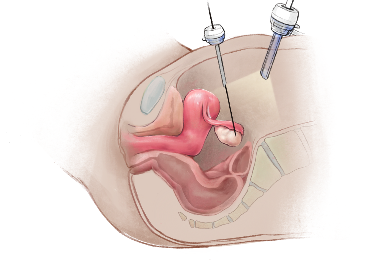
Figure 1.2a: Sagittal cross-section showing a laparoscopic oocyte retrieval.
Despite this novel milestone, the laparoscopic technique had limitations. Retrieving only one oocyte at a time, the need for general anesthesia and an operating room, and longer procedure time limited success rates. Lenz and colleagues 7 were the first to demonstrate the feasibility of transabdominal ultrasound-guided oocyte retrieval (TAOR). This was further refined by Gleicher and colleagues, as well as Dellenbach and colleagues, to develop transvaginal ultrasound-guided oocyte retrieval (TVOR). Unlike previous techniques, TVOR could be performed in an outpatient setting without general anesthesia. Currently, most IVF centers globally perform oocyte retrievals using ultrasound-guided techniques, with TVOR being the preferred route. 2
1.3 TVOR vs. TAOR
TVOR and TAOR are briefly discussed in the following table. 8
| Technique | Transvaginal (TVOR) | Transabdominal (TAOR) |
|---|---|---|
| Tools used to visualize ovaries | Transvaginal ultrasound transducer with detachable needle guide | May use an abdominal transducer, or the same setup as TVOR |
| Image |
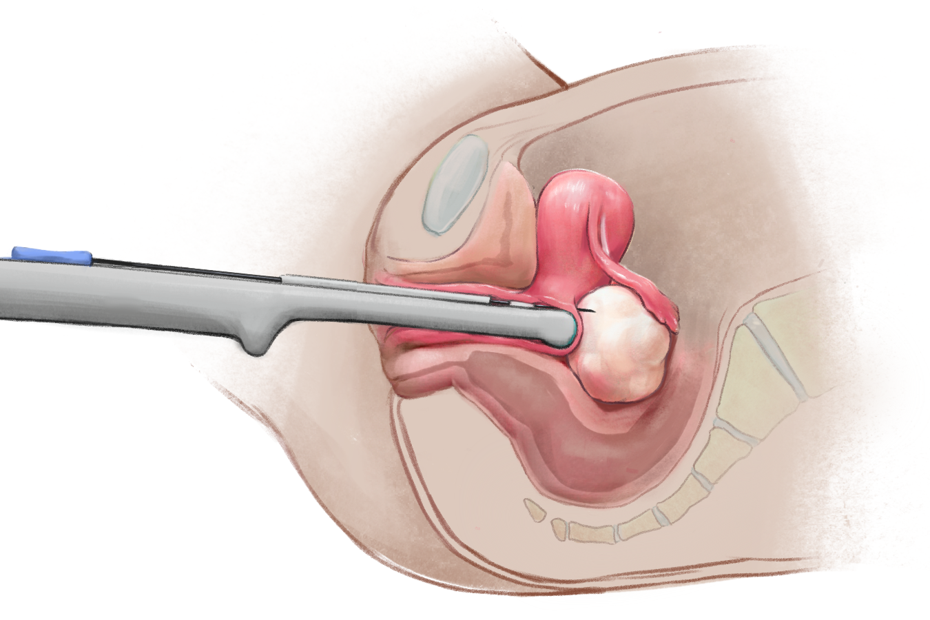
Figure 1.3a: Sagittal cross-section showing a transvaginal oocyte retrieval. |
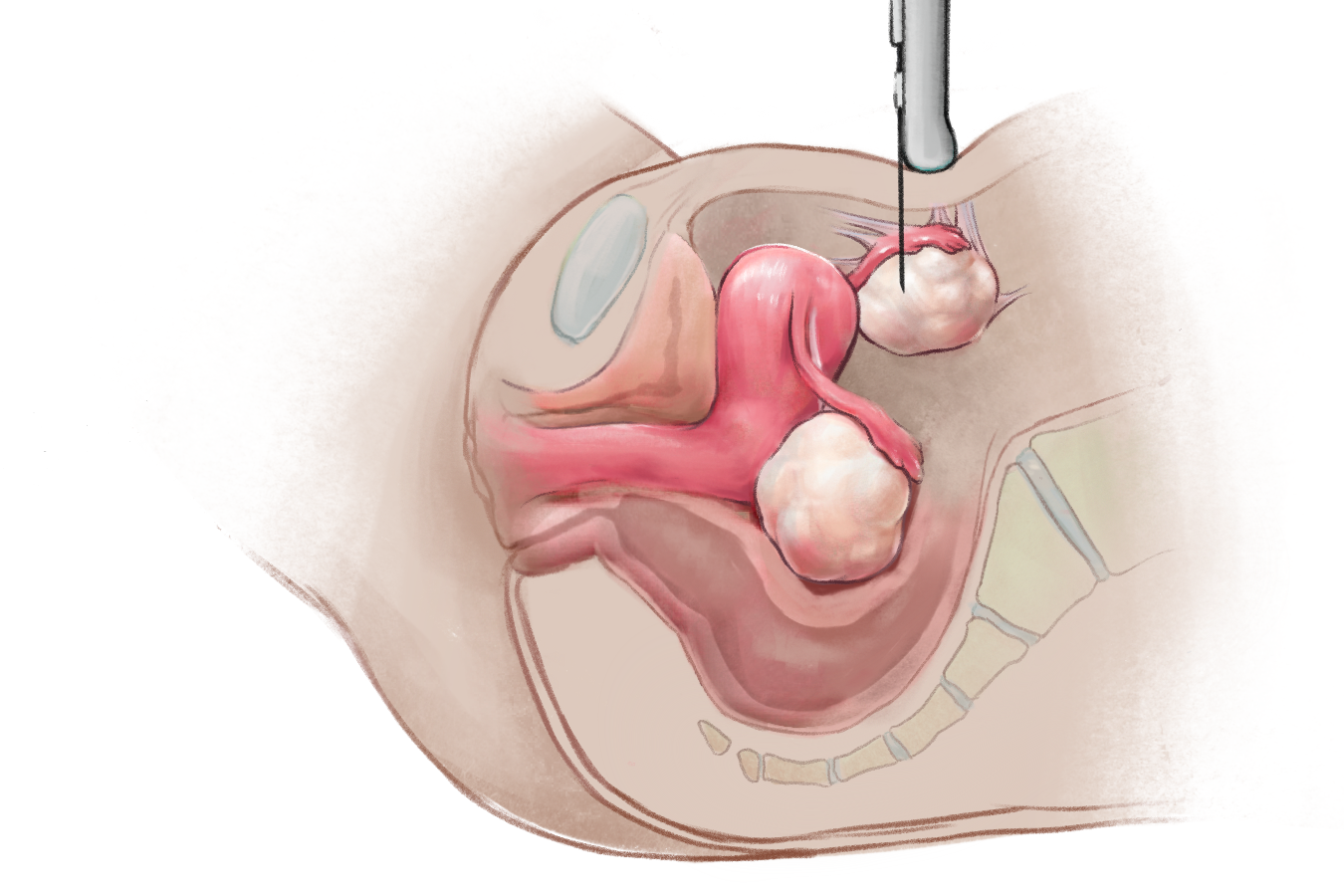
Figure 1.3b: Sagittal cross-section showing a transabdominal oocyte retrieval. |
| Structures punctured | Needle passes through the posterior fornix of the vaginal canal to reach the ovarian follicles | Needle passes through the anterior abdominal wall to reach the ovarian follicles |
| Sedation | Local anesthesia and conscious sedation | Local anesthesia and conscious sedation OR general anesthesia |
| Follicle aspiration | External pump connected to aspiration needle | External pump connected to aspiration needle |
| Notes |
Preferred technique
|
Technique used in patients whose ovaries cannot be accessed transvaginally. Indications may include: |
1.4 Indications and contraindications
The most common indications for oocyte retrieval are:
- Collection of oocytes for IVF
- Planned oocyte cryopreservation
- Urgent fertility preservation
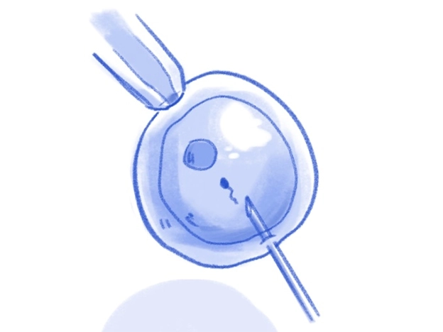
Figure 1.4a: Collection of oocytes for IVF

Figure 1.4b: Planned oocyte cryopreservation
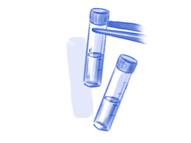
Figure 1.4c: Urgent fertility preservation
The TVOR set-up can also be used for the aspiration of ovarian cysts, hydrosalpinges, or ovarian hyperstimulation-related paracentesis, as well as transmyometrial embryo transfer.
While a detailed list of contraindications remains beyond the scope of this guide, TVOR and TAOR should be avoided in clinical situations associated with increased risk of bleeding, infection, injury or tumor seeding in the abdominopelvic cavity. These include but are not limited to:2
- Active abdominopelvic infection
- Uncontrolled or undiagnosed bleeding or clotting disorders
- Abdominopelvic masses suspicious for malignancy

Figure 1.4d: Active abdominopelvic infection
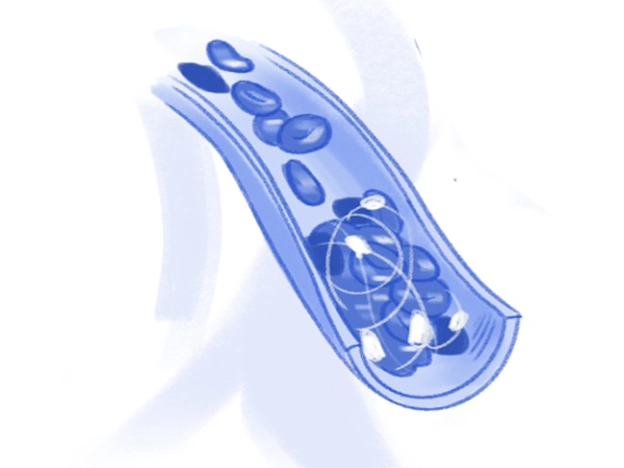
Figure 1.4e: Uncontrolled or undiagnosed bleeding or clotting disorders
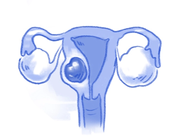
Figure 1.4f: Abdominopelvic masses suspicious for malignancy
References
- Adamson GD, Creighton P, de Mouzon J, Zegers-Hochschild F, Dyer S, Chambers GM. How many infants have been born with the help of assisted reproductive technology? Fertil Steril. 2025;124:40-50. Available from: https://doi.org/10.1016/j.fertnstert.2025.02.009
- Pereira N, Fitz VW. Complications of oocyte retrieval. In: Lindheim SR, Petrozza JC, editors. Reproductive surgery. 1st ed. Cham: Springer; 2022. p. 287-98. Available from: https://doi.org/10.1007/978-3-031-05240-8_23
- Rock J, Menkin MF. In vitro fertilization and cleavage of human ovarian eggs. Science. 1944;100(2588):105-7. Available from: https://doi.org/10.1126/science.100.2588.105
- Menkin MF, Rock J. In vitro fertilization and cleavage of human ovarian eggs. Am J Obstet Gynecol. 1948;55(3):440-52. Available from: https://www.ajog.org/article/S0002-9378(15)32963-X/abstract
- Steptoe PC. Laparoscopy and ovulation. Lancet. 1968;2(7574):913. Available from: https://www.thelancet.com/journals/lancet/article/PIIS0140-6736(68)91079-9/fulltext
- Healy MW, Hill MJ, Levens ED. Optimal oocyte retrieval and embryo transfer techniques: where we are and how we got here. Semin Reprod Med. 2015;33(2):83-91. Available from: https://doi.org/10.1055/s-0035-1545365
- Lenz S, Lauritsen JG, Kjellow M. Collection of human oocytes for in vitro fertilisation by ultrasonically guided follicular puncture. Lancet. 1981;1(8230):1163-4. Available from: https://www.thelancet.com/journals/lancet/article/PIIS0140-6736(81)92335-7/fulltext
- D'Angelo A, Panayotidis C, Amso N, et al.; ESHRE Working Group on Ultrasound in ART. Recommendations for good practice in ultrasound: oocyte pick up. Hum Reprod Open. 2019;2019(4):hoz025. Available from: https://doi.org/10.1093/hropen/hoz025
- Shenoy CC, Coddington C, Khan Z, Jones TL, Jensen J. Experience in transabdominal oocyte retrievals: a case series. Fertil Steril. 2016;106(3). Available from: https://www.fertstert.org/article/S0015-0282(16)61456-5/fulltext
- Pereira N, Willson S, McCarter K, Chung PH, Kligman I, Rosenwaks Z. Case report: interventional radiology's potential role for in vitro fertilization post ovarian transposition and pelvic radiation. Fertil Steril. 2021;115(5):1347-9. Available from: https://doi.org/10.1016/j.fertnstert.2020.12.006